拡散反射分光法に基づく生体医用光学

メンバー: 西舘泉
分野: 電気電子工学、人間医工学、応用物理学
所属: 工学研究院 先端電気電子部門
キーワード: 生体医用光学、 組織分光学
ウェブサイト:
研究概要
私たちの体は大きさや機能の異なる多くの細胞により構成され、メラニンやヘモグロビンなどに代表される、生命活動に不可欠ないくつかの色素蛋白を含んでいます。例えば皮膚に光を当てると、その一部は体内に入り込み、細胞組織や色素により散乱、吸収されます。この生体と光の相互作用は組織構造や色素の種類、状態により異なる分光(スペクトル)特性を示すことが知られています。この生体の分光特性を積極的に利用することで生体組織の機能情報を非侵襲的に計測し、医療診断・治療に役立てようとする試みが広くなされており、そのような研究分野は生体医用光学と呼ばれています。
生体医用光学の分野において、様々な光学的手法が研究・開発されています。中でも、拡散光を利用した分光計測は、丸ごとの生体や生きた細胞組織から得られる吸収・散乱スペクトル情報を解析することで生体の機能及び構造情報を評価することができることから、様々な応用が期待できます。とりわけ、定常白色光源を用いた拡散反射分光法(Diffuse reflectance spectroscopy, DRS)は簡易・安価な計測システムで実現可能であり、in vivo測定やイメージングへの展開も容易であるという利点を持っています。
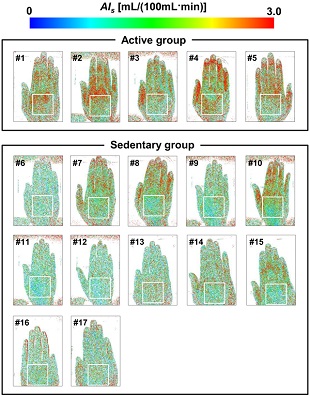
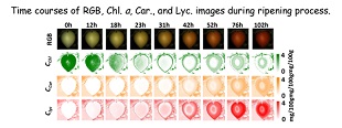
当研究グループでは主に可視波長域の拡散光スペクトル及び色彩情報に基づき、生体内の生理的変化に対する色素タンパク質(メラニン、ヘモグロビン)の挙動や細胞組織の構造形態変化をin vivoで評価するための方法について、理論的および実験的検討を行っています。また、ヒトや動物を対象とした検討に加え、植物や作物を対象とした非破壊計測・機能イメージングに関する検討も進めており、食糧問題や環境問題など、植物育成、増産を促進する新しい技術の創出へ向けた農工融合研究も積極的に進めています。
主要論文・参考事項
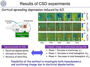
1) I. Nishidate, C. Mizushima, K. Yoshida, S. Kawauchi, S. Sato, and M. Sato, “In vivo estimation of light scattering and absorption properties of rat brain using a single-reflectance fiber probe during cortical spreading depression,” Journal of Biomedical Optics, Vol.20, No.2, 027003-1-027003-14 (2015).
2) K. Yoshida, I. Nishidate, T. Ishizuka, S. Kawauchi, S. Sato, and M. Sato, “Multispectral imaging of absorption and scattering properties of in vivo exposed rat brain using a digital red-green-blue camera,” Journal of Biomedical Optics, Vol.20, No.5, 051026-1-051026-15 (2015).
3) K. Yoshida and I. Nishidate, “Rapid calculation of diffuse reflectance from a multilayered model by combination of the white Monte Carlo and adding-doubling methods,” Biomedical Optics Express, Vol.5, pp.3901-3920 (2014).
4) S. Sato, S. Kawauchi, W. Okuda, I. Nishidate, H. Nawashiro, G. Tsumatori, “Real-Time Optical Diagnosis of the Rat Brain Exposed to a Laser-Induced Shock Wave: Observation of Spreading Depolarization, Vasoconstriction and Hypoxemia-Oligemia,” PLoS ONE, Vol. 9, No. 1, pp. e82891-1-e82891-15 (2014).
5) I. Nishidate, N. Tanaka, T. Kawase, T. Maeda, T. Yuasa, Y. Aizu, T. Yuasa, K. Niizeki, “Visualization of peripheral vasodilative indices in human skin by use of Red, Green, Blue images,” Journal of Biomedical Optics, Vol.18, No.6, pp.061220-1-061220-9 (2013).
6) I. Nishidate, T. Maeda, K. Niizeki, Y. Aizu, “Estimation of melanin and hemoglobin using spectral reflectance images reconstructed from a digital RGB image by Wiener estimation method,” Sensors, Vol.13, No.6, 7902-7915 (2013).
7) I. Nishidate, N. Tanaka, T. Kawase, T. Maeda, T. Yuasa, Y. Aizu, T. Yuasa, and K. Niizeki, “Noninvasive imaging of human skin hemodynamics using a digital red-green-blue camera,” Journal of Biomedical Optics, Vol.16, No.8, pp. 086012-1- 086012-14 (2011).
お問い合わせ先
東京農工大学・先端産学連携研究推進センター
urac[at]ml.tuat.ac.jp([at]を@に変換してください)
Biomedical optics based on diffuse reflectance spectroscopy
Research members: Dr. Izumi Nishidate
Research fields: Electrical and electronic engineering, Biomedical engineering, Applied physics
Departments: Institute of Engineering, Division of Advanced Electrical and Electronics Engineering
Keywords: Biomedical Optics, Tissue spectroscopy
Web site:
Summary
In the field of biomedical optics, the optical properties of biological tissue have been used to evaluate spatial and/or temporal changes of physiological and morphological conditions in living tissues. The changes in optical properties of the tissue occur mainly by the following processes: changes in absorption and scattering originating from hemodynamics, changes in absorption due to redox states of cytochromes in mitochondria, changes in scattering generated by cell swelling or shrinkage caused by water movement between intracellular and extracellular compartments, and changes in scattering and absorption caused by chromophore contents and cell deformations. Light in the visible to near-infrared spectral range is sensitive to the absorption and scattering properties of biological tissue. Tissue optics has become the established general term for techniques that deal with the optical properties of biological tissues.
Tissue optics combines the optical technologies with biological and medical sciences to advance healthcare treatment, including diagnosis, monitoring, treatment and therapy. In recent years, the tissue optics for clinical diagnostics and therapeutic medicine has been attracted as the non- and minimally-invasive technique and, thus, various optical methods have been studied. Some biological chromophores and scatterers are related to the physiological and pathophysiological conditions of tissue, and have their own absorption and scattering spectra. Light scattered by biological tissues has interacted with the ultrastructure of the tissue. Ultrastructure of tissue extends from membranes to membrane aggregates to collagen fibers to nuclei to cells. The light are most strongly scattered by those structures whose size matches its wavelength. A number of spectroscopic methods have been studied for in vivo determination of the scattering and absorption properties in living tissues, including time-resolved measurements, a frequency-domain method, and spatially resolved measurements. Diffuse reflectance spectroscopy (DRS) with continuous wave light can be simply achieved with a white light source, inexpensive optical components, and a spectrometer. Multispectral imaging is a useful tool for extension of DRS to evaluate the spatial maps of tissue optical properties. Multispectral imaging can be simply achieved with a monochromatic CCD camera with narrow-band filters and a white light source, and has been employed to monitor tissue oxygenation, distinction between normal and malignant tissue, and assessment of tissue viability.
Biomedical optics field consists of a foundation in physics, chemistry, mathematics, biology, physiology and medicine for engineers to design optical instrumentation for biomedical applications. This field of study has a large potential in the study of various problems in the life sciences ranging from molecular level researches to clinical treatment of patients and animals.
Reference articles and patents
1) I. Nishidate, C. Mizushima, K. Yoshida, S. Kawauchi, S. Sato, and M. Sato, “In vivo estimation of light scattering and absorption properties of rat brain using a single-reflectance fiber probe during cortical spreading depression,” Journal of Biomedical Optics, Vol.20, No.2, 027003-1-027003-14 (2015).
2) K. Yoshida, I. Nishidate, T. Ishizuka, S. Kawauchi, S. Sato, and M. Sato, “Multispectral imaging of absorption and scattering properties of in vivo exposed rat brain using a digital red-green-blue camera,” Journal of Biomedical Optics, Vol.20, No.5, 051026-1-051026-15 (2015).
3) K. Yoshida and I. Nishidate, “Rapid calculation of diffuse reflectance from a multilayered model by combination of the white Monte Carlo and adding-doubling methods,” Biomedical Optics Express, Vol.5, pp.3901-3920 (2014).
4) S. Sato, S. Kawauchi, W. Okuda, I. Nishidate, H. Nawashiro, G. Tsumatori, “Real-Time Optical Diagnosis of the Rat Brain Exposed to a Laser-Induced Shock Wave: Observation of Spreading Depolarization, Vasoconstriction and Hypoxemia-Oligemia,” PLoS ONE, Vol. 9, No. 1, pp. e82891-1-e82891-15 (2014).
5) I. Nishidate, N. Tanaka, T. Kawase, T. Maeda, T. Yuasa, Y. Aizu, T. Yuasa, K. Niizeki, “Visualization of peripheral vasodilative indices in human skin by use of Red, Green, Blue images,” Journal of Biomedical Optics, Vol.18, No.6, pp.061220-1-061220-9 (2013).
6) I. Nishidate, T. Maeda, K. Niizeki, Y. Aizu, “Estimation of melanin and hemoglobin using spectral reflectance images reconstructed from a digital RGB image by Wiener estimation method,” Sensors, Vol.13, No.6, 7902-7915 (2013).
7) I. Nishidate, N. Tanaka, T. Kawase, T. Maeda, T. Yuasa, Y. Aizu, T. Yuasa, and K. Niizeki, “Noninvasive imaging of human skin hemodynamics using a digital red-green-blue camera,” Journal of Biomedical Optics, Vol.16, No.8, pp. 086012-1- 086012-14 (2011).
Contact
University Research Administration Center(URAC),
Tokyo University of Agriculture andTechnology
urac[at]ml.tuat.ac.jp
(Please replace [at] with @.)