原子間力顕微鏡技術を用いた木材細胞壁ミクロフィブリル傾角測定システムの開発

メンバー: 佐藤敬一
分野: 森林圏科学、材料工学、生体分子科学
所属: 農学研究院 農学部環境資源科学科
キーワード: 原子間力顕微鏡、セルロースミクロフィブリル、仮道管細胞壁
研究概要
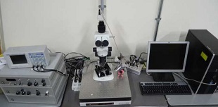
原子間力顕微鏡のミクロフィブリル傾角等の木材物性評価への応用可能性を検討するために、まず、原子間力顕微鏡(FM検出方式)を用いてヒノキ仮道管壁縦断面を観察した。
飽水状態のヒノキ木片からスライディング・ミクロトームを用いて板目切削面、15°切削面、板目切片、15°切片、30°切片の5種類の試料を作成し、シリカゲル入りのデシケータ内で乾燥させた後、仮道管壁縦断面を観察した。
各条件において、長径150~600 nm、短径100~300 nm 程度の楕円形の構造が壁一面に認められる視野があった。15°切削面、板目切片、30°切片の3条件では直径100~300 nmの円形の構造が認められる視野があった。スライディング・ミクロトームを用いた切削や、飽水処理と乾燥処理による非結晶領域内の水溶性成分の除去や細胞壁の膨潤・収縮によって楕円形や円形の構造の存在が強調されたために、観察された可能性がある。ミクロフィブリルの束の横断面が円形であると仮定すると、断面形状は傾角が小さいほど楕円形に、傾角が大きいほど円形に近い形状になると考えられる。観察された楕円形や円形の構造がミクロフィブリルの束に由来するものであれば、そこに傾角等の情報が含まれると考えられ、傾角測定の検討が期待できる。しかし、楕円や円形の構造はすべての視野では認められなかったので、まずこれらの構造を安定的に観察できる条件を検討する必要がある。
主要論文・参考事項
「分子間力プローブ顕微鏡技術を用いた細胞壁ミクロフィブリル傾角測定システムの開発」平成26年度科学研究費補助事業(挑戦的萌芽研究)研究成果報告書
お問い合わせ先
東京農工大学・先端産学連携研究推進センター
urac[at]ml.tuat.ac.jp([at]を@に変換してください)
Development of measurement system with atomic force microscope for cell wall microfibril angle
Research members: Keiichi Sato PhD.
Research fields: Forest and forest products science, Material engineering, Biomolecular science
Departments: Department of Environment and Natural Resouces, Institute of Agriculture
Keywords: atomic force microscope, cellose microfibril, cell wall of tracheid
Summary
By using Atomic Force Microscope, cell walls of tracheid (Chamaecyparis obtusa) were observed to find the possibility of Atomic Force Microscope for evaluation of wood physical characteristics such as microfibril angle.
The oval shaped structure(longer axis:150~600 nm, minor axis:100~300 nm) was observed on tracheid on each sumples. On 15°cutting side, thin section of cross gain, 30°thin section, we observed round structure (diamieter:100~300 nm). Cutting by sliding microtome, and removal of water soluble component in non-crystalline dmain and swelling-shrincage by water satulation and drying might lead to observation of oval shaped structure and round structure. If bundle of microfibrils has round shaped cross-section, it has oval shape at small microfibril angle, and has round shape at greater microfibril angle. We guess that if oval shape and round shape are derive from bundle of microfibrils, these contain information on microfibril angle, we may be able to measure microfibril angles. But, we have to investigate the condtion that can observe these structure stably.
Reference articles and patents
「分子間力プローブ顕微鏡技術を用いた細胞壁ミクロフィブリル傾角測定システムの開発」平成26年度科学研究費補助事業(挑戦的萌芽研究)研究成果報告書
Contact
University Research Administration Center(URAC),
Tokyo University of Agriculture andTechnology
urac[at]ml.tuat.ac.jp
(Please replace [at] with @.)