Development of measurement system with atomic force microscope for cell wall microfibril angle

Research members: Keiichi Sato PhD.
Research fields: Forest and forest products science, Material engineering, Biomolecular science
Departments: Department of Environment and Natural Resouces, Institute of Agriculture
Keywords: atomic force microscope, cellose microfibril, cell wall of tracheid
Summary
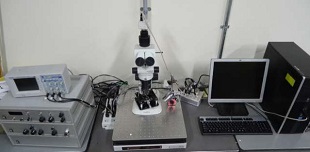
By using Atomic Force Microscope, cell walls of tracheid (Chamaecyparis obtusa) were observed to find the possibility of Atomic Force Microscope for evaluation of wood physical characteristics such as microfibril angle.
The oval shaped structure(longer axis:150~600 nm, minor axis:100~300 nm) was observed on tracheid on each sumples. On 15°cutting side, thin section of cross gain, 30°thin section, we observed round structure (diamieter:100~300 nm). Cutting by sliding microtome, and removal of water soluble component in non-crystalline dmain and swelling-shrincage by water satulation and drying might lead to observation of oval shaped structure and round structure. If bundle of microfibrils has round shaped cross-section, it has oval shape at small microfibril angle, and has round shape at greater microfibril angle. We guess that if oval shape and round shape are derive from bundle of microfibrils, these contain information on microfibril angle, we may be able to measure microfibril angles. But, we have to investigate the condtion that can observe these structure stably.
Reference articles and patents
「分子間力プローブ顕微鏡技術を用いた細胞壁ミクロフィブリル傾角測定システムの開発」平成26年度科学研究費補助事業(挑戦的萌芽研究)研究成果報告書
Contact
University Research Administration Center(URAC),
Tokyo University of Agriculture andTechnology
urac[at]ml.tuat.ac.jp
(Please replace [at] with @.)